


自迁移出 MediaWiki 以来,本页内容尚未经过审查。如果您愿意帮忙,请查看帮助指南!
引文
请注意,斐济提供的 SPIM 注册插件基于书籍。如果您成功将其用于您的研究,请引用我们的工作:
- S.普雷比什、S.萨尔菲尔德、J. Schindelin 和 P. Tomancak (2010)“用于流动平面照明工作站数据的基于珠的配准的软件”,《自然方法》,7(6):418-419。 Webpage PDF Supplement
重要提示
有关 SPIM 注册、融合和反格式的详细信息,请查看Multiview Reconstruction Plugin。它更加强大、灵活并且与BigDataViewer集成完全。有关过时的SPIM Registration的文档仍然可用。
简介
SPIM 原则
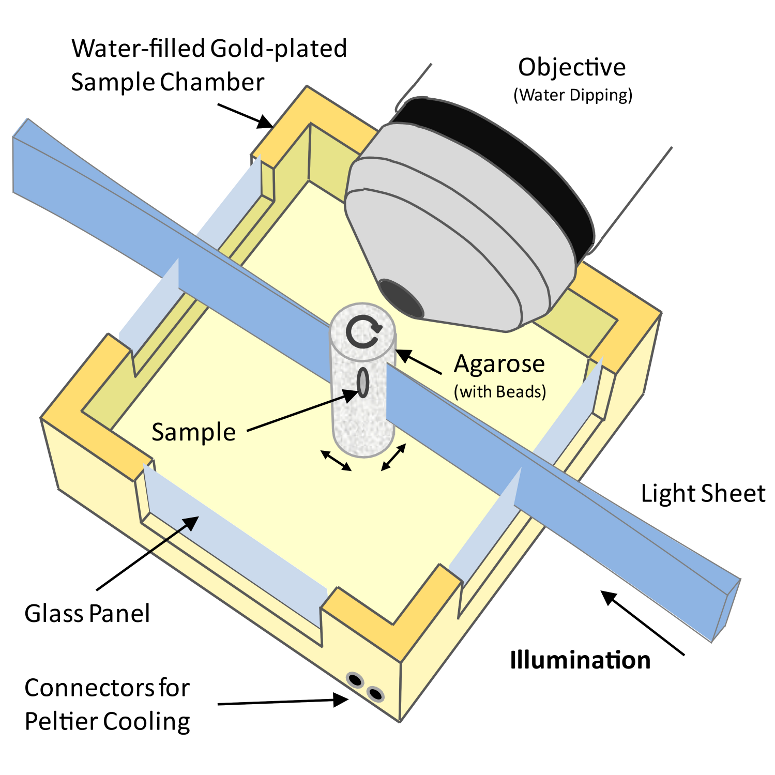 Figure 1: Schematic drawing of a Selective Plane Illumination Microscope
A Selective Plane Illumination Microscope[^1] (Figure 1), achieves optical sectioning by focusing the excitation laser into a thin laser light sheet that reaches its minimal thickness in the middle of the field of view. The light sheet enters the water filled sample chamber and illuminates the sample which is embedded in an agarose column. The agarose protrudes from the end of a glass capillary attached to a motor that rotates the sample. The objective lens is arranged perpendicular to the light sheet. The perpendicular orientation of the illumination and detection optics ensures that only a section of the specimen in-focus is illuminated, minimizing photo-bleaching and laser damage of the living samples and allowing for very long time-lapse recordings. Two-dimensional images of emitted fluorescent light are captured by a CCD camera focussed on the center of the light-sheet. The CCD camera captures the light-sheet-illuminated section in a single exposure enabling a very fast acquisition rate important for capturing dynamic developmental events. In order to acquire 3d image stacks, the sample is moved through the light sheet in increments of 0.5 μm to 5 μm depending on the objective and the light sheet thickness.
原则上,SPIM 仪器可以沿着 \(x\)、\(y\) 和 \(z\) 轴实现各向同性的高分辨率,从而允许对大型 3D 样本进行“整体”成像。为了在所有三个维度上在样本体积上实现均匀的各向同性分辨率,有必要旋转样本并从不同角度记录同一样本的图像堆栈(通常为 3 到 12 个,参见Video 1)。
###相关工作
多视图显微技术,例如倾斜共焦采集、[^2] SPIM[^3] 和超显微术[^4](2 个视图),可以提高沿 \(z\) 轴的分辨率,从而能够分析小于轴向分辨率限制的物体。[^5][^6] 基于图像强度的图像重建需要图像内容的显着重叠,这通常很难实现,特别是在动态变化样本的实时成像中。
合并基准标记以促进样本独立重建的想法广泛应用于医学成像[^7][^8][^9][^10][^11]和电子断层扫描。[^12][^13]由于可用于配准的基准标记数量很少,因此研究重点是误差分析,而不是仅部分重叠的数千个标记的匹配效率。[^14]
相比之下,在机器人和自动化领域,人们对大量不同物体的定位感兴趣。从照片中提取兴趣点,并对照数据库进行检查以确定其类型和方向。[^15][^16]为了实现实时对象识别,Lamdan 等人[^17]引入了“几何哈希”,它使用固有不变的局部坐标系以独立于视点的方式将对象与数据库条目进行匹配。几何哈希原理在天文学[^18]和蛋白质结构比对和比较[^19][^20][^21][^22]领域中得到重用,这些领域需要在大量点云中进行高效搜索。
在图像配准、[^23][^24]机器人和自治系统、[^25][^26]和计算机视觉等许多领域中都提出使用局部描述符而不是完整场景进行匹配。[^27]
Matula 等人[^28]提出了基于分割的方法来重建多视图显微图像。分段对象云的质心用作柱坐标系的参考点,有助于两个视图之间的配准。与基于强度的方法类似,该方法需要图像之间存在大量重叠,并且一次仅支持两个堆栈的对齐。
我们的方法结合了使用信托标记、局部描述符和几何哈希的思想,并应用了全局优化。它可以注册任意数量的部分重叠的点云。它在合并的珠子数量、珠子分布、重叠量方面具有鲁棒性,并且可以可靠地检测成像过程中可能发生的非仿射干扰(例如突然的琼脂糖运动)(Table 1)。
方法
珠子分割
合并的亚分辨率珠子在每个图像 \(I(x,y,z)\) 中显示为微观系统的点扩散函数 (PSF)。为了检测珠子,理想情况下将其与显微镜的脉冲响应 (PSF) 进行卷积,从而在每个珠子的亚像素位置产生最高的相关性。然而,由于曝光时间、激光功率、珠子类型、物镜和琼脂糖浓度的变化,PSF 在不同的实验中并不是恒定的。此外,由于光片的凹度,PSF 在整个视场中并不是恒定的,因此卷积运算的计算量非常大。 我们发现,适当平滑的 3d LaPlace 滤波器 \(\nabla^2\) 能够以足够的精度检测所有珠子,同时有效抑制高频噪声。正如计算机视觉文献中所建议的,[^29][^30] 我们通过图像 \(I\) 的两个高斯卷积 (DoG) 的差异来近似 \(\nabla^2I\),标准差 \(\sigma\) 分别为 1.4 px 和 1.8 px。 \(\nabla^2I\) 中 3×3×3 邻域中的所有局部最小值表示强度最大值,然后通过将 3d 二次函数拟合到该邻域来估计其子像素位置。[^31] DoG 检测器可以识别珠子,即使它们彼此靠近、靠近样本或具有意外形状的珠子。它还对图像进行大规模过度分割,检测成像样本内的“斑点状”结构、角落以及沿边缘或平面的各个位置。然而,这些检测不会干扰注册过程,因为包含它们的描述符被本地描述符匹配过滤掉(参见Figure 2)。在不同的视图中仅重复检测到珠子。
建立珠子对应关系
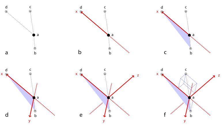 Figure 2: Rotation invariant local geometric descriptor
To register two views \(A\) and \(B\) the corresponding bead pairs \((\vec{a},\vec{b})\) have to be identified invariantly to translation and rotation. To this end, we developed a geometric local descriptor . The local descriptor of a bead is defined by the locations of its 3 nearest neighbors in 3d image space ordered by their distance to the bead. To efficiently extract the nearest neighbors in image space we use the kd-tree implementation of the WEKA framework.[^32] Translation invariance is achieved by storing locations relative to the bead. That is, each bead descriptor is an ordered 3d point cloud of cardinality 3 with its origin \(\vec{o} = (0,0,0)^T\)} being the location of the bead.
通过使用基于闭式单位四元数的解决方案,通过最小二乘点映射误差将所有珠子 \(\vec{a}\in A\) 的有序点云分别映射到所有珠子 \(\vec{b}\in B\) 的有序点云,从而对旋转不变地执行局部描述符匹配。[^33] 相似性度量 \(\epsilon\) 是平均点映射误差。 \(A\) 中的每个候选者都与 \(B\) 中的每个候选者匹配。相应的描述符是那些具有最小 \(\epsilon\) 的描述符。然而,这种方法的计算要求非常高,因为它的检测数量复杂度为 \(O(n^2)\)。[^34]
因此,我们采用了几何哈希的变体[^35]来加速匹配过程。我们没有为整个场景使用一个参考坐标系,而是为每个描述符定义一个局部坐标系,如第 Figure 2 中所示和所述。所有未用于定义局部坐标系的剩余珠子坐标都变得旋转不变,这使我们能够使用 kd 树非常有效地比较描述符,以索引局部坐标系中的剩余珠子坐标。再次,我们在六维树上采用 WEKA 框架 [^36] 的 kd-tree 实现来识别描述符空间中的最近邻居,即最相似的描述符。明显优于描述符空间中第二个最近邻的最相似描述符被指定为对应候选者。[^37]
仅由四个珠子组成的描述符并不完全独特,并且类似的描述符可能会偶然出现。增加描述符中的珠子数量将使其更加独特,但代价是较少识别的对应关系和增加的计算时间。所有真实对应都同意一种转换模型以实现最佳视图配准,而每个错误对应支持不同的转换。因此,我们使用最小描述符大小(4 个珠子),并在仿射变换模型上使用随机样本共识 (RANSAC)[^38] 拒绝候选集的错误对应,然后进行稳健回归。
全局优化
已识别的一组相应珠子 \(C_{AB} = \{(\vec{a}_i,\vec{b}_i) : i=\{1,2, \dots, \left| C\right|\}\}\) 对于一对视图 \(A\) 和 \(B\) 定义仿射变换 \(\mathbf{T}_{AB}\),通过最小二乘珠对应位移将 \(A\) 映射到 \(B\)
我们使用仿射变换来校正由水和琼脂糖之间的差系数不匹配引入的各向异性\(z\)拉伸[^39][^40],因为样品永远不会完美地位于琼脂糖柱的中心。
注册两个以上视图需要对配置进行分组优化 \(T_{VF} = \{\mathbf{T}_{AF} : A,F\in V\}\),其中 \(V\) 是所有视图的集合,\(F\) 是定义公共参考系的固定视图。那么,上式延伸为
\(C_{AB}\) 是视图 \(A\) 和视图 \(B\) 之间的珠对应集 \((\vec{a},\vec{b})\),而 \(\vec{a}\ 在 A\) 和 \(\vec{b}\ 在 B\) 中。该项是使用迭代优化方案求解的。在每次迭代中,估计 V\setminus{F}\(中每个单个视图\)A\ 相对于所有其他视图的当前配置的最佳仿射变换 \(\mathbf{T}_{AF}\) 并将其应用于该视图中的所有珠子。该方案在整体珠对应位移收敛时终止。该解决方案允许我们在显微镜设置具有不同属性(例如平移[^41]、刚性[^42])的情况下使用任何转换模型执行全局优化。
延时注册
在长时间延时成像期间,整个琼脂糖柱可能会移动。为了补偿漂移,我们使用基于珠子的配准框架来相互配准各个时间点。我们从系列中间的任意时间点\(t\)中选择单个视图\(A_t\)作为参考。随后,我们使用存储的 DoG 检测来识别所有视图对 \(A_t\) 和 \(A_{a\in{\{1,2, \dots \}\setminus\{t\}}}\) 的真实对应局部几何描述符,并计算仿射变换 \(\mathbf{T}_{A_aA_t}\),通过最小二乘珠将 \(A_a\) 映射到 \(A_t\)对应位移。然后,将识别的变换矩阵应用于相应时间点的所有剩余视图,从而形成注册的时间序列(Figure 4a)。
图像融合和混合
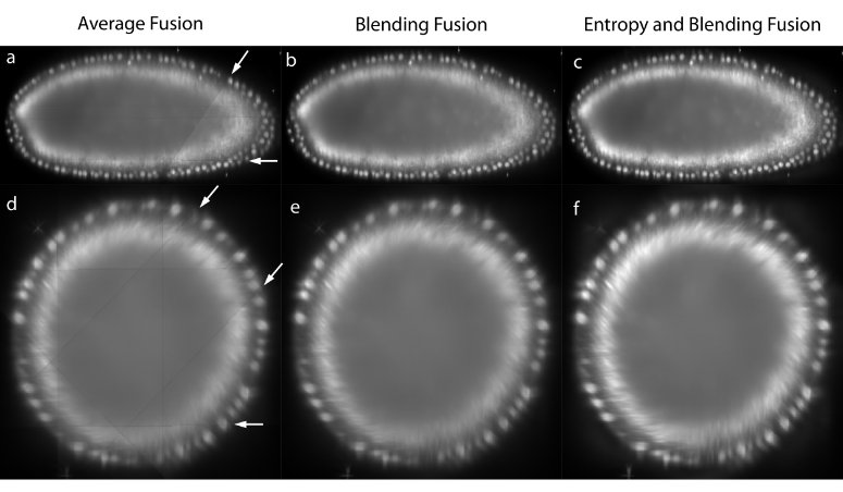 Figure 3: Image fusion
The registered views can be combined to create a single isotropic 3d image. An effective fusion algorithm must ensure that each view contributes to the final fused volume only useful sharp image data acquired from the area of the sample close to the detection lens. Blurred data visible in other overlapping views should be suppressed. We use Gaussian Filters to approximate the image information at each pixel in the contributing views (Figure 3c,f).[^43]
对于像果蝇这样的强散射和吸收样本,我们通常不会在每个单一视图中对整个样本进行成像,而是随着图像变得越来越模糊和扭曲而停止在其深度的大约三分之二处。在重建的 3D 图像中,这会在视图突然结束的区域引入线条伪影 (Figure 3a,d)。为了抑制这种影响以实现数据显示,我们在靠近视图之间每个边框的边缘处应用非线性混合 (Figure 3b,e)。[^44]
多视图数据的精确配准是重建图像多视图反卷积的先决条件,这可能会提高分辨率。[^45][^46][^47] 在样本周围具有亚分辨率荧光珠有助于估计空间相关的点扩散函数并验证反卷积结果。
珠子去除策略
用于注册视图的亚分辨率荧光珠的存在可能会干扰数据集的后续分析。为了通过计算从每个视图中移除珠子,我们通过添加相应视图的所有真实对应项(用于配准的珠子)的局部图像邻域来计算平均珠子形状。获得的模板随后用于识别其他珠子;为了加快检测速度,我们仅将此模板与初始珠子分割步骤期间高斯差分算子检测到的所有最大值进行比较。这些 DoG 检测包含所有图像最大值,因此包含样本的所有珠子。然后通过减去珠子模板的标准化、高斯模糊版本来去除珠子。该方法可靠地去除了通过检测到的珠子附近的平均强度判断的与样品明显分离的珠子。因此,一些非常靠近样品的珠子不会被去除,因为珠子的扣除会干扰样品的强度。
为了从样品中完全去除所有珠子,我们根据成像样品调整珠子的强度。因此,我们只需将可被不同波长激发的珠子嵌入样品中,然后使用长通滤光片进行检测(Figure 6e,g)。在这种采集中,珠子的强度约为 2-4。
评价
基于珠子的配准框架的性能评估
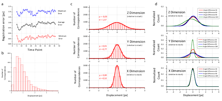 Figure 4: Analysis of the registration error
We created a visualization of the optimization procedure. For each view, we display its bounding box and the locations of all corresponding descriptors in a 3d visualization framework[^48]. Correspondences are color coded logarithmically according to their current displacement ranging from red (>100 px) to green (<1 px). The optimization is initialized with a configuration where the orientation of the views is unknown; all views are placed on top of each other and thus the corresponding descriptor displacement is high (red). As the optimization proceeds, the average displacement decreases (yellow) until convergence at about one pixel average displacement (green) is achieved. Video 2 shows the optimization progress for an 8 angle acquisition of fixed C.elegans . The outline of the worm forms in the middle (grey), since many worm nuclei were segmented by the DoG detector but discarded during establishment of bead correspondences. 全局优化方案可以无缝应用于平铺多视图采集。在这样的设置中,使用高倍率镜头从多个角度分别扫描大型 3D 样品的不同部分。所有此类采集都可以混合,丢弃有关其排列的所有信息,并且全局优化恢复正确的配置。 Video 3中显示了这种优化的一个例子,其中包含一个固定的果蝇胚胎,在带有40×/0.8 Achroplan物镜的单光子共焦显微镜上从每个角度的两个或三个瓦片的8个角度成像。
为了证明基于珠子的配准框架的准确性,我们创建了一个模拟的仅珠子数据集,其中的珠子由 σ=1.5 px 的高斯滤波器响应近似。我们生成了 8 个不同的视图,这些视图通过具有各向同性分辨率的近似刚性仿射变换相关。该数据集的重建产生了 0.02 px (Table 1) 的平均误差。对于现实数据集,配准通常会导致约 1 px 或稍低的误差(Figure 4b 和 Table 1),其中剩余误差是由珠检测器的定位精度和琼脂糖弹性变形引起的小非仿射干扰引入的。在每个视图的 \(xy\) 平面中,可以非常精确地定位珠子,但是,与 \(z\) 一起,由于采样率较低和 PSF 不对称,定位精度会下降。第 Figure 4c,d 中显示的尺寸相关误差支持这一点。
Table 1表明,对于配准,我们通常使用比解决仿射变换所需的更多的珠子(4个珠子)。需要使用很多珠子进行注册,原因如下:
- 如果堆栈之间的重叠(如许多示例所示)非常小,则必须确保在这些小的重叠区域中仍然有足够的基准标记来对齐堆栈。
- 如果两个堆栈广泛重叠,则珠子必须均匀分布在样品周围,以确保整个样品的误差分布均匀。否则,例如左下角的小误差无法控制没有基准标记的右上角的配准误差。
- 珠子的定位误差呈正态分布,如 Figure 4c,d 所示。这意味着,注册中包含的珠子越多,珠子的平均定位就越准确。因此,使用的珠越多,每个单独堆栈的仿射变换产生的残余误差就越低。
由于样品的光学特性,珠子可能会发生扭曲和偏差,从而在错误的位置进行检测。通常,此类珠子被排除在外,因为它们不会在不同堆栈之间形成可重复的描述符。如果畸变很小,它们将导致仿射变换的残余误差。然而,贡献很小,因为这些珠子只占所有珠子的很小一部分。仿射模型 1 px 的最大传输误差支持这一点(另请参见第 Figure 5c,d 的插图)。
Video 4和Video 5中显示了在时间点内和跨时间点登记的果蝇胚胎发育时间序列的示例。视频显示了所有细胞中表达 His-YFP 的发育胚胎的 3D 效果图。从四个和三个任意角度显示 3D 渲染的单个胚胎,以突出标本的完整覆盖。 Video 4 以 42 个时间点(7 个角度)显示了原肠胚形成后的最后两次同步核分裂。 Video 5 在 249 个时间点(5 个角度)果蝇 胚胎发生过程中捕获从原肠胚形成到成熟胚胎的过程,此时肌肉活动有效地阻止了进一步成像。
基于珠子的配准框架与基于强度的方法的性能比较
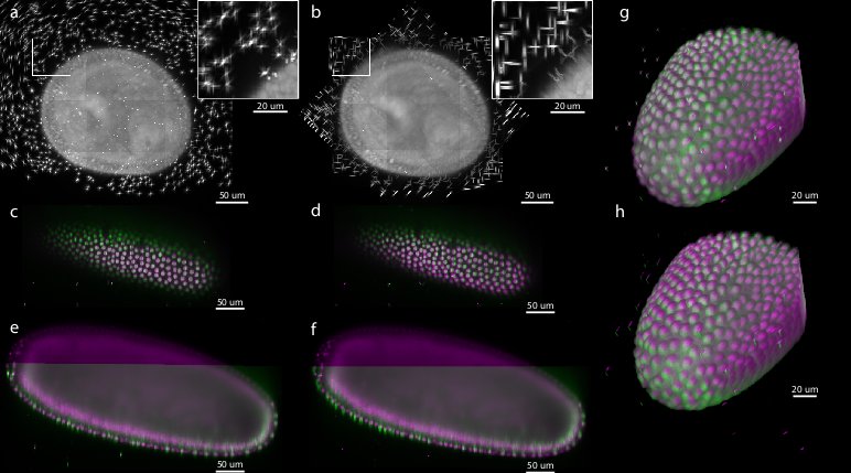 Figure 5: Comparison of bead-based and intensity-based multi-view reconstruction on 7-view acquisition of Drosophila embryo expressing His-YFP
Existing muti-view SPIM registration approaches that use sample intensities to iteratively optimize the quality of the overlap of the views do not work reliably and are computationally demanding.[^49][^50][^51] Alternatively, the registration can be achieved by matching of segmented structures, such as cell nuclei, between views[^52] However, such approaches are not universally applicable, as the segmentation process has to be adapted to the imaged sample. 为了评估基于珠子的配准框架的精度和性能,我们将其与我们之前开发的基于强度的配准方法进行了比较。[^53]该方法通过相邻视图之间基于 FFT 的相位相关性的迭代优化来识别所有视图公共的旋转轴。我们将这两种方法(基于珠子和基于强度)应用于单个时间点实时 7 视图采集果蝇胚胎,该胚胎在嵌入带有珠子的琼脂糖中的所有细胞中表达 His-YFP。我们选择了胚盘阶段的一个时间点,其中胚胎的形态随时间变化最小。我们通过相应珠子的平均位移评估了这两种方法的精度,并得出结论,在珠子配准精度方面,基于珠子的配准框架明显优于基于强度的配准(0.98 px vs 6.91 px,参见Table 1、Figure 5)。基于珠子的配准框架所实现的珠子对准精度的提高反映在样品中细胞核重叠的显着改善(参见Figure 5c–h)。此外,基于强度的方法需要大约 9 小时的计算时间,而在相同计算机硬件(具有 64GB RAM 的 Intel Xeon E5440)上执行的基于珠子的配准框架则需要 2.5 分钟,也就是说,基于珠子的框架对于该数据集大约快 200 倍。 ————————————————————————————————
|
数据集 |
最小/平均/最大托盘 [px] |
狗检测 |
对应真实数(比例) |
处理时间[分:秒] |
|---|---|---|---|---|
|
固定的线虫, 8次观看 |
1.02/1.12/1.31 |
4566 |
1717 (98%) |
11:09 |
|
居住果蝇, 5次浏览 |
0.76/0.81/1.31 |
9267 |
1459 (97%) |
2:31 |
|
固定的果蝇, 10次浏览 |
0.65/0.78/0.97 |
9035 |
1301 (93%) |
20:10 |
|
固定的果蝇, 11次浏览 |
1.10/1.33/1.86 |
6309 |
978 (92%) |
6:15 |
|
模拟数据集,8个视图 |
0.02/0.02/0.02 |
2594 |
2880 (96%) |
15:54 |
|
居住果蝇, 7次浏览 |
0.87/0.98/1.17 |
6232 |
603 (97%) |
2:27 |
|
居住果蝇, 7次浏览 |
0.93/6.91/9.59 |
n.a. |
n.a. |
515:10 |
样式=“边距顶部:1em;边距底部:1em;” |表1: 各数据集多视图配准统计
我们展示了全局优化收敛后所有真实对应(珠子)的最小、平均和最大位移。模拟数据集显示配准错误非常低。尽管 DoG 检测可以参与多个对应对,但 DoG 检测的总数通常远高于提取的对应候选对的数量。真实对应与对应候选人的比例通常高于 90%。较低的比率表明存在配准问题,例如由堆栈采集期间琼脂糖的移动引起的。处理时间(分段和配准)是在双四核 Intel Xeon E5440 系统上测量的。
SPIM 成像的样本
成像样本概述
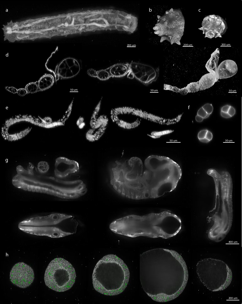 Figure 6: Examples of various reconstructed multi-view datasets
We demonstrated the performance of our registration framework on multi-view in toto imaging of fixed and living specimen of various model organisms (Figure 6), in particular Drosophila . Fixed Drosophila embryos were stained with Sytox-Green to label all nuclei. For live imaging, we used a developing Drosophila embryo expressing fluorescent His-YFP under the control of endogenous promoter visualizing all nuclei. Drosophila specimens were imaged with a SPIM prototype equipped with a Zeiss 20×/0.5 Achroplan objective.
SPIM 样品安装
我们将珠子的荧光强度与样品的信号强度相匹配。对于相对暗淡且需要较长曝光时间(0.3 秒)的 His-YFP 实时成像,我们使用红色或黄色荧光珠,这些珠子对于 GFP 检测滤光片组来说不是最佳的,因此通常不如样品明亮。相反,对于明亮、固定样本的成像,我们使用绿色荧光珠,它可以在非常短的曝光时间(0.01 秒)下提供足够的信号。
尽管我们的算法在可用于注册的珠子数量方面是稳健的,但过多的珠子不必要地增加了计算时间,而太少的珠子可能由于视图不完全重叠而导致对应数量不足。因此,我们根据经验确定了每个放大倍数的最佳珠子浓度(理想情况下每个成像体积有 1,000–2,000 个珠子)。我们制备了 2× 珠子储备溶液(13 μl 浓缩珠子溶液(Estapor 微球 FXC050))
独立注册示例
我们将基于珠子的注册框架应用于来自主要模式生物的各种样品。其中包括果蝇胚胎、幼虫 (Figure 6a–c) 和卵子发生 (Figure 6d),C。线虫成体(数据未显示)、幼虫阶段(Figure 6e)和早期胚胎(Figure 6f)、整个小鼠胚胎(Figure 6g)和斑马鱼胚胎的双色成像(Figure 6h)。尽管样品的大小、荧光标记、光学特性和安装格式差异很大,但基于珠子的配准框架始终能够实现配准。因此,我们得出的结论是,我们的方法是独立于样本的,并且普遍适用于任何多视图 SPIM 采集的注册,其中样本移动不会干扰琼脂糖的刚性。
基于珠子的框架对多视图成像的广泛适用性
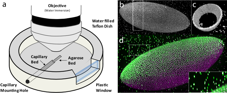 Figure 7: Multi-view imaging with spinning disc confocal microscopy
Having the bead-based registration framework for multi-view reconstruction established, we sought to expand its application beyond SPIM, to other microscopy techniques capable of multi-view acquisition.[^54] We designed a sample-mounting set-up that allows imaging of a sample embedded in a horizontally positioned agarose column with fluorescent beads (Figure 7a). The agarose column was manually rotated mimicking the SPIM multi-view acquisition. We acquired multiple views of fixed Drosophila embryos stained with nuclear dye on a spinning disc confocal microscope and reconstructed the views using the bead-based registration framework. By mosaicking around the sample, we captured the specimen in toto and achieved full lateral resolution in areas that are compromised by the poor axial resolution of a single-view confocal stack (Figure 7b,c,d and Video 6 ). The combination of multi-view acquisition and bead-based registration is applicable to any imaging modality as long as the fluorescent beads can be localized and the views overlap.
在正置显微镜上进行多视图成像的样品安装
我们构建了一个用于在正置显微镜上进行多视图成像的样品室,该样品室由侧壁上配有孔的聚四氟乙烯盘组成,该孔的直径与标准玻璃毛细管 (Figure 7a) 相同。毛细管安装孔在培养皿底部继续延伸,形成半圆形沟槽,厚度约为毛细管直径的一半,向培养皿中心延伸约培养皿半径的 2/3。该沟槽用作通过毛细管安装孔插入的玻璃毛细管的床。该沟槽由第二个较浅的沟槽延伸,该沟槽的底部相对于较深的沟槽升高了毛细管玻璃壁的厚度。第二个沟槽用作琼脂糖柱的床,琼脂糖柱通过紧密配合的柱塞(未示出)被推出毛细管。聚四氟乙烯盘的一侧配备有塑料窗口,可以目视检查样品和物镜。
为了成像,将样品嵌入含有适量荧光珠的琼脂糖中的毛细管插入毛细管安装孔,直到到达毛细管床的末端。聚四氟乙烯培养皿装满水,柱塞将琼脂糖从毛细管中推出到琼脂糖床中。将水浸物镜放入培养皿中,并聚焦在琼脂糖中的果蝇胚胎样本上。使用各种光学切片技术(旋转圆盘共焦(§§2§§§)、单光子共焦(参见Video 3)、双光子共焦、apotome(数据未显示))获得共焦堆栈。接下来,为了实现多视图采集,通过柱塞将琼脂糖柱缩回到毛细管中,并手动旋转毛细管。旋转角度仅通过所附胶带片的位置非常粗略地估计琼脂糖再次被推入琼脂糖床中,并以这种方式收集任意数量的视图,只要样品不漂白。
实施
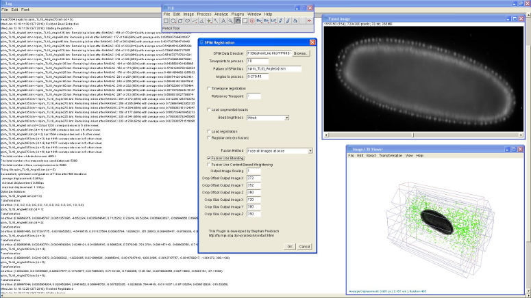 Figure 8: Screenshot of SPIM registration plugin in Fiji
The bead-based registration framework is implemented in the Java programming language and provided as a fully open source plugin packaged with the ImageJ distribution Fiji (Fiji Is Just ImageJ, that is actively developed by an international group of developers. The plugin (Figure 8) performs all steps of the registration pipeline: bead segmentation, correspondence analysis of bead-descriptors, outlier removal (RANSAC and global regression), global optimization including optional visualization, several methods for fusion, blending and time-lapse registration.
有关如何在基本和高级模式下使用该插件的教程,请参阅SPIM Registration。包含 果蝇 胚胎 7 视图 SPIM 采集的测试数据可以从 [^1](http://fly.mpi-cbg.de/preibisch/nm/HisYFP-SPIM.zip) 下载。
致谢
我们要感谢 Carl Zeiss Microimaging 访问 SPIM 演示者、His-YFP 苍蝇的 Radoslav Kamil Ejsmont[^55]、Dan White、[^56]、Jonathan Rodenfels、[^57] Ivana Viktorinova、[^58] Mihail Sarov、[^59] Steffen Jänsch、[^60] Jeremy Pulvers、[^61] 和Pedro Campinho[^62] 提供各种生物样本以使用 Figure 6 中所示的 SPIM 进行成像。
参考文献
[^1]:<div class="notice notice-default" style="font-size: 4;"><div class="notice-icon">❌</div><div class="notice-content"><p>Invalid citation arguments: {"content"=>"journal", "author"=>"P. Shaw, D. Agard, Y. Hiraoka, and J. Sedat", "title"=>"Tilted view reconstruction in optical microscopy. Three-dimensional reconstruction of Drosophila melanogaster embryo nuclei", "journal"=>"Biophysical Journal", "volume"=>"55", "number"=>"1", "year"=>"1989", "pages"=>"101–110"}</p>
</div>
</div>
[^2]:<div class="notice notice-default" style="font-size: 4;"><div class="notice-icon">❌</div><div class="notice-content"><p>Invalid citation arguments: {"content"=>"journal", "title"=>"Optical Sectioning Deep Inside Live Embryos by Selective Plane Illumination Microscopy", "journal"=>"Science", "volume"=>"305", "number"=>"5686", "pages"=>"1007 1010", "year"=>"2004"}</p>
</div>
</div>
[^3]:
[^4]:<div class="notice notice-default" style="font-size: 4;"><div class="notice-icon">❌</div><div class="notice-content"><p>Invalid citation arguments: {"content"=>"journal", "author"=>"J. Swoger, P. Verveer, K. Greger, J. Huisken, and E. H. K. Stelzer", "journal"=>"Opt. Express", "number"=>"13", "pages"=>"8029–8042", "title"=>"Multi-view image fusion improves resolution in three-dimensional microscopy", "volume"=>"15", "year"=>"2007"}</p>
</div>
</div>
[^5]:
[^7]:<div class="notice notice-default" style="font-size: 4;"><div class="notice-icon">❌</div><div class="notice-content"><p>Invalid citation arguments: {"content"=>"journal", "author"=>"B.J. Erickson and Jack C.R. Jr.", "title"=>"Correlation of single photon emission CT with MR image data using fiduciary markers", "journal"=>"American Journal of Neuroradiology", "volume"=>"14", "number"=>"3", "pages"=>"713 720", "year"=>"1993"}</p>
</div>
</div>
[^8]:<div class="notice notice-default" style="font-size: 4;"><div class="notice-icon">❌</div><div class="notice-content"><p>Invalid citation arguments: {"title"=>"Three Dimensional X-Ray Opaque Foreign Body Marker Device", "author"=>"E. H. Gullekson", "year"=>"Patent number: 3836776, Filing date: 1 Mar 1973, Issue date: Sep 1974"}</p>
</div>
</div>
[^9]:<div class="notice notice-default" style="font-size: 4;"><div class="notice-icon">❌</div><div class="notice-content"><p>Invalid citation arguments: {"content"=>"journal", "author"=>"A. D. Wiles, A. Likholyot, D. D. Frantz, and T.M.Peters", "title"=>"A Statistical Model for Point-Based Target Registration Error With Anisotropic Fiducial Localizer Error", "journal"=>"IEEE Transactions on Medical Imaging", "volume"=>"27", "number"=>"3", "year"=>"2008", "month"=>"March", "pages"=>"378 390"}</p>
</div>
</div>
[^10]:<div class="notice notice-default" style="font-size: 4;"><div class="notice-icon">❌</div><div class="notice-content"><p>Invalid citation arguments: {"content"=>"journal", "author"=>"J. M. Fitzpatrick and J. B. West", "title"=>"The distribution of target registration error in rigid-body point-based registration", "journal"=>"IEEE Transactions on Medical Imaging", "volume"=>"20", "number"=>"9", "year"=>"2001", "month"=>"September", "pages"=>"917 927"}</p>
</div>
</div>
[^11]:<div class="notice notice-default" style="font-size: 4;"><div class="notice-icon">❌</div><div class="notice-content"><p>Invalid citation arguments: {"content"=>"journal", "title"=>"Towards automatic electron tomography", "journal"=>"Ultramicroscopy", "volume"=>"40", "number"=>"1", "pages"=>"71 87", "year"=>"1992", "author"=>"K. Dierksen, D. Typke, R. Hegerl, A. J. Koster, and W. Baumeister"}</p>
</div>
</div>
[^12]:doi:10.1007/978-3-540-85990-1_124
[^13]:<div class="notice notice-default" style="font-size: 4;"><div class="notice-icon">❌</div><div class="notice-content"><p>Invalid citation arguments: {"content"=>"conference", "title"=>"A Combined Corner and Edge Detector", "author"=>"C. Harris and M. Stephens", "locaiton"=>"Plessey Research Roke Manor, UK", "booktitle"=>"Proceedings of The Fourth Alvey Vision Conference", "location"=>"Manchester", "year"=>"1988", "pages"=>"147 151"}</p>
</div>
</div>
[^14]:
[^15]:<div class="notice notice-default" style="font-size: 4;"><div class="notice-icon">❌</div><div class="notice-content"><p>Invalid citation arguments: {"content"=>"journal", "author"=>"A. J. Koster, R. Grimm, D. Typke, R. Hegerl, A. Stoschek, J. Walz, and W. Baumeister", "title"=>"Perspectives of molecular and cellular electron tomography", "journal"=>"J Struct Biol", "year"=>"1997", "volume"=>"120", "number"=>"3", "pages"=>"276 308"}</p>
</div>
</div>
[^16]:<div class="notice notice-default" style="font-size: 4;"><div class="notice-icon">❌</div><div class="notice-content"><p>Invalid citation arguments: {"content"=>"conference", "author"=>"Y. Lamdan, J. Schwartz, and H. Wolfson", "title"=>"On recognition of 3D objects from 2D images", "booktitle"=>"Proceedings of the IEEE International Conference on Robotics and Automation", "year"=>"1988", "pages"=>"1407 1413", "publisher"=>"IEEE Computer Society", "location"=>"Los Alamitos, CA"}</p>
</div>
</div>
[^17]:<div class="notice notice-default" style="font-size: 4;"><div class="notice-icon">❌</div><div class="notice-content"><p>Invalid citation arguments: {"content"=>"journal", "title"=>"A computational approach to edge detection", "author"=>"J. Canny", "journal"=>"IEEE Transactions on Pattern Analysis Machine Intelligence", "volume"=>"8", "number"=>"6", "year"=>"1986", "pages"=>"679 698"}</p>
</div>
</div>
[^18]:<div class="notice notice-default" style="font-size: 4;"><div class="notice-icon">❌</div><div class="notice-content"><p>Invalid citation arguments: {"content"=>"journal", "author"=>"R. Nussinov and H. J. Wolfson", "journal"=>"Proc Natl Acad Sci U S A", "number"=>"23", "pages"=>"10495 10499", "title"=>"Efficient detection of three-dimensional structural motifs in biological macromolecules by computer vision techniques.", "volume"=>"88", "year"=>"1991"}</p>
</div>
</div>
[^19]:<div class="notice notice-default" style="font-size: 4;"><div class="notice-icon">❌</div><div class="notice-content"><p>Invalid citation arguments: {"content"=>"conference", "author"=>"D. W. Hogg, M. Blanton, D. Lang, K. Mierle, and S. Roweis", "title"=>"Automated Astrometry", "booktitle"=>"Astronomical Data Analysis Software and Systems XVII", "year"=>"2008", "series"=>"Astronomical Society of the Pacific Conference Series", "volume"=>"394", "editor"=>"R. W. Argyle, P. S. Bunclark, and J. R. Lewis", "month"=>"August", "pages"=>"27-+"}</p>
</div>
</div>
[^20]:<div class="notice notice-default" style="font-size: 4;"><div class="notice-icon">❌</div><div class="notice-content"><p>Invalid citation arguments: {"content"=>"journal", "author"=>"A.C. Wallace, N. Borkakoti, and J. M. Thornton", "title"=>"Tess: A geometric hashing algorithm for deriving 3D coordinate templates for searching structural databases. Application to enzyme active sites", "journal"=>"Protein science", "year"=>"1997", "volume"=>"6", "number"=>"11", "pages"=>"2308"}</p>
</div>
</div>
[^21]:<div class="notice notice-default" style="font-size: 4;"><div class="notice-icon">❌</div><div class="notice-content"><p>Invalid citation arguments: {"content"=>"journal", "author"=>"D. Fischer, H. Wolfson, S. L. Lin, and R. Nussinov", "title"=>"Three-dimensional, sequence order-independent structural comparison of a serine protease against the crystallographic database reveals active site similarities: Potential implications to evolution and to protein folding", "journal"=>"Protein Science", "volume"=>"3", "number"=>"5", "pages"=>"769 778", "year"=>"1994"}</p>
</div>
</div>
[^22]:<div class="notice notice-default" style="font-size: 4;"><div class="notice-icon">❌</div><div class="notice-content"><p>Invalid citation arguments: {"content"=>"conference", "author"=>"A. Stanski and O. Hellwich", "title"=>"Spiders as Robust Point Descriptors", "booktitle"=>"DAGM-Symposium", "year"=>"2005", "pages"=>"262 268"}</p>
</div>
</div>
[^23]:<div class="notice notice-default" style="font-size: 4;"><div class="notice-icon">❌</div><div class="notice-content"><p>Invalid citation arguments: {"content"=>"journal", "title"=>"A Model for Statistical Significance of Local Similarities in Structure", "journal"=>"Journal of Molecular Biology", "volume"=>"326", "number"=>"5", "pages"=>"1307 1316", "year"=>"2003", "author"=>"A. Stark, S. Sunyaev, and R. B. Russell"}</p>
</div>
</div>
[^24]:<div class="notice notice-default" style="font-size: 4;"><div class="notice-icon">❌</div><div class="notice-content"><p>Invalid citation arguments: {"content"=>"journal", "author"=>"B. Kuipers and Yung-tai Byun", "title"=>"A Robot Exploration and Mapping Strategy Based on a Semantic Hierarchy of Spatial Representations", "journal"=>"Journal of Robotics and Autonomous Systems", "year"=>"1991", "volume"=>"8", "pages"=>"47 63"}</p>
</div>
</div>
[^25]:<div class="notice notice-default" style="font-size: 4;"><div class="notice-icon">❌</div><div class="notice-content"><p>Invalid citation arguments: {"content"=>"journal", "author"=>"D. G. Lowe", "title"=>"Distinctive image features from scale-invariant keypoints", "journal"=>"Int J Comput Vis", "volume"=>"60", "number"=>"2", "pages"=>"91 110", "year"=>"2004"}</p>
</div>
</div>
[^26]:<div class="notice notice-default" style="font-size: 4;"><div class="notice-icon">❌</div><div class="notice-content"><p>Invalid citation arguments: {"content"=>"conference", "author"=>"A. Frome, D. Huber, R. Kolluri, T. Bulow, and J. Malik", "title"=>"Recognizing objects in range data using regional point descriptors", "booktitle"=>"Proceedings of the European Conference on Computer Vision (ECCV)", "month"=>"May", "year"=>"2004"}</p>
</div>
</div>
[^27]:<div class="notice notice-default" style="font-size: 4;"><div class="notice-icon">❌</div><div class="notice-content"><p>Invalid citation arguments: {"content"=>"conference", "author"=>"D. Bradley, D. Silver, and S. Thayer", "title"=>"A regional point descriptor for global localization in subterranean environments", "booktitle"=>"IEEE conference on Robotics Automation and Mechatronics (RAM 2005)", "pages"=>"440 445", "month"=>"December", "year"=>"2004", "volume"=>"1"}</p>
</div>
</div>
[^28]:<div class="notice notice-default" style="font-size: 4;"><div class="notice-icon">❌</div><div class="notice-content"><p>Invalid citation arguments: {"content"=>"journal", "author"=>"T. Lindeberg", "title"=>"Scale-space theory: A basic tool for analysing structures at different scales", "journal"=>"Journal of Applied Statistics", "volume"=>"21", "number"=>"2", "pages"=>"224 270", "year"=>"1994"}</p>
</div>
</div>
[^29]:<div class="notice notice-default" style="font-size: 4;"><div class="notice-icon">❌</div><div class="notice-content"><p>Invalid citation arguments: {"content"=>"journal", "author"=>"P. Matula, M. Kozubek, F. Staier, and M. Hausmann", "journal"=>"Journal of microscopy", "pages"=>"126 42", "title"=>"Precise 3D image alignment in micro-axial tomography.", "volume"=>"209", "year"=>"2003"}</p>
</div>
</div>
[^30]:
[^31]:<div class="notice notice-default" style="font-size: 4;"><div class="notice-icon">❌</div><div class="notice-content"><p>Invalid citation arguments: {"content"=>"book", "author"=>"I. H. Witten and E. Frank", "title"=>"Data Mining: Practical machine learning tools and techniques", "publisher"=>"Morgen Kaufmann", "location"=>"San Francisco", "isbn"=>"0-12-088407-0", "edition"=>"second", "year"=>"2005"}</p>
</div>
</div>
[^32]:<div class="notice notice-default" style="font-size: 4;"><div class="notice-icon">❌</div><div class="notice-content"><p>Invalid citation arguments: {"content"=>"conference", "author"=>"M. Brown and D. Lowe", "title"=>"Invariant Features from Interest Point Groups", "booktitle"=>"In British Machine Vision Conference", "year"=>"2002", "pages"=>"656 665"}</p>
</div>
</div>
[^33]:doi:10.1117/12.812612
[^34]:<div class="notice notice-default" style="font-size: 4;"><div class="notice-icon">❌</div><div class="notice-content"><p>Invalid citation arguments: {"content"=>"journal", "author"=>"B. K. P. Horn", "title"=>"Closed-form solution of absolute orientation using unit quaternions", "journal"=>"Journal of the Optical Society of America A", "volume"=>"4", "number"=>"4", "pages"=>"629 642", "year"=>"1987"}</p>
</div>
</div>
[^35]:
[^36]:
[^37]:
[^38]:<div class="notice notice-default" style="font-size: 4;"><div class="notice-icon">❌</div><div class="notice-content"><p>Invalid citation arguments: {"content"=>"journal", "title"=>"Aberrations in Confocal Fluorescence Microscopy Induced by Mismatches in Refractive Index", "journal"=>"Journal of Microscopy", "volume"=>"169", "number"=>"3", "pages"=>"341 405", "year"=>"1993", "author"=>"S. Hell and G. Reiner and C. Cremer and E.H.K. Stelzer"}</p>
</div>
</div>
[^39]:<div class="notice notice-default" style="font-size: 4;"><div class="notice-icon">❌</div><div class="notice-content"><p>Invalid citation arguments: {"content"=>"journal", "author"=>"M. A. Fischler and R. C. Bolles", "title"=>"Random sample consensus: a paradigm for model fitting with applications to image analysis and automated cartography", "journal"=>"Communications of the ACM", "volume"=>"24", "number"=>"6", "pages"=>"381 395", "year"=>"1981"}</p>
</div>
</div>
[^40]:
[^41]:doi:10.1117/12.770893
[^42]:
[^43]:doi:10.1093/bioinformatics/btp184
[^44]:
[^45]:
[^46]:<div class="notice notice-default" style="font-size: 4;"><div class="notice-icon">❌</div><div class="notice-content"><p>Invalid citation arguments: {"content"=>"journal", "title"=>"High-resolution three-dimensional imaging of large specimens with light sheet-based microscopy", "journal"=>"Nature Methods", "volume"=>"4", "number"=>"4", "pages"=>"311 313", "year"=>"2007", "author"=>"P.J. Verveer and J. Swoger and F. Pampaloni and K. Greger and M. Marcello and E.H.K.Stelzer"}</p>
</div>
</div>
[^47]:<div class="notice notice-default" style="font-size: 4;"><div class="notice-icon">❌</div><div class="notice-content"><p>Invalid citation arguments: {"content"=>"journal", "author"=>"C. J. Engelbrecht and E. H. K. Stelzer", "title"=>"Resolution enhancement in a light-sheet-based microscope (SPIM)", "journal"=>"Optics Letters", "volume"=>"31", "number"=>"10", "pages"=>"1477 1479", "year"=>"2006"}</p>
</div>
</div>
[^48]:<div class="notice notice-default" style="font-size: 4;"><div class="notice-icon">❌</div><div class="notice-content"><p>Invalid citation arguments: {"content"=>"journal", "author"=>"Jim Swoger and Peter Verveer and Klaus Greger and Jan Huisken and Ernst H. K. Stelzer", "journal"=>"Opt. Express", "number"=>"13", "pages"=>"8029 8042", "publisher"=>"OSA", "title"=>"Multi-view image fusion improves resolution in three-dimensional microscopy", "volume"=>"15", "year"=>"2007"}</p>
</div>
</div>
[^49]:<div class="notice notice-default" style="font-size: 4;"><div class="notice-icon">❌</div><div class="notice-content"><p>Invalid citation arguments: {"content"=>"conference", "author"=>"B. Schmidt", "title"=>"Hardware-accelerated 3D visualization for ImageJ", "booktitle"=>"ImageJ User and Developer Conference", "year"=>"2008", "volume"=>"2", "editor"=>"Pierre Plumer"}</p>
</div>
</div>
[^50]:<div class="notice notice-default" style="font-size: 4;"><div class="notice-icon">❌</div><div class="notice-content"><p>Invalid citation arguments: {"content"=>"conference", "author"=>"S. Preibisch and R. Ejsmont and T. Rohlfing and P. Tomancak", "title"=>"Towards Digital Representation of Drosophila Embryogenesis", "booktitle"=>"Proceedings of 5th IEEE International Symposium on Biomedical Imaging", "pages"=>"324 327", "year"=>"2008"}</p>
</div>
</div>
[^51]:doi:10.1117/12.770893
[^52]:<div class="notice notice-default" style="font-size: 4;"><div class="notice-icon">❌</div><div class="notice-content"><p>Invalid citation arguments: {"content"=>"conference", "author"=>"S. Preibisch and R. Ejsmont and T. Rohlfing and P. Tomancak", "title"=>"Towards Digital Representation of Drosophila Embryogenesis", "booktitle"=>"Proceedings of 5th IEEE International Symposium on Biomedical Imaging", "pages"=>"324 327", "year"=>"2008"}</p>
</div>
</div>
[^53]:<div class="notice notice-default" style="font-size: 4;"><div class="notice-icon">❌</div><div class="notice-content"><p>Invalid citation arguments: {"content"=>"journal", "author"=>"P. J. Keller and A. D. Schmidt and J. Wittbrodt and E. H. Stelzer", "title"=>"Reconstruction of zebrafish early embryonic development by scanned light sheet microscopy", "journal"=>"Science", "volume"=>"322", "number"=>"5904", "pages"=>"1065-9", "year"=>"2008"}</p>
</div>
</div>
[^54]:<div class="notice notice-default" style="font-size: 4;"><div class="notice-icon">❌</div><div class="notice-content"><p>Invalid citation arguments: {"content"=>"journal", "author"=>"J. Bradl and M. Hausmann and V. Ehemann and D. Komitowski and C. Cremer", "title"=>"A tilting device for three-dimensional microscopy: application to in situ imaging of interphase cell nuclei.", "journal"=>"Journal of Microscopy", "year"=>"1992", "volume"=>"168", "pages"=>"47 57"}</p>
</div>
</div>
[^55]:马克斯·普朗克分子细胞生物学和遗传学研究所,德累斯顿,德国
[^56]:
[^57]:
[^58]:
[^59]:
[^60]:
[^61]:
[^62]: